Congenital Pulmonary Airway Malformation (CPAM) in Pregnancy: Causes, Ultrasound Diagnosis, Risks, and Treatment
Finding an abnormality in your baby’s lungs during a prenatal ultrasound can be frightening, particularly when the condition has a medical name that you have never heard before. One such condition is Congenital Pulmonary Airway Malformation (CPAM), a rare developmental abnormality of the fetal lung.
CPAM was previously called congenital cystic adenomatoid malformation (CCAM). It occurs when abnormal lung tissue develops during pregnancy and forms a mass that does not function normally. CPAM is usually detected during a routine prenatal ultrasound, often during the second trimester.
Although the diagnosis can be concerning, many babies with CPAM have a favorable outcome, particularly when the lesion remains small and does not interfere with normal lung development or fetal circulation.
For pregnant women in the Philippines, understanding CPAM, how it is monitored, and when specialist care may be necessary can help families prepare for the remainder of pregnancy and delivery.
What Is Congenital Pulmonary Airway Malformation?
Congenital Pulmonary Airway Malformation is a developmental abnormality in which a portion of the baby’s lung develops abnormally.
The affected area may contain:
- Small or large fluid-filled cysts
- Multiple cysts
- Solid-appearing abnormal lung tissue
- A combination of cystic and solid areas
CPAM usually affects one lung and is most commonly confined to a single lobe. The abnormal tissue does not function like normal lung tissue.
Most CPAMs are benign, meaning they are not cancerous.
The condition develops while the baby’s lungs are forming and is generally considered a sporadic developmental abnormality rather than something inherited from the parents.
What Causes CPAM?
The exact cause of CPAM is not known.
It occurs because of abnormal development of the fetal airways and lung tissue during pregnancy. There is currently no evidence that a mother’s normal activities, diet, exercise, stress, or routine behavior causes CPAM.
CPAM is generally considered a random developmental event and is not usually inherited.
Parents should therefore not blame themselves if their baby is diagnosed with CPAM.
How Common Is CPAM?
CPAM is considered a rare congenital lung abnormality. Modern ultrasound technology has made it possible to identify many cases before birth that previously may not have been diagnosed until after delivery.
A 2024 review from the Philippine Children’s Medical Center examined 23 CPAM cases treated between 2011 and 2021. In that series, only four patients, or 17.39%, had CPAM detected prenatally by ultrasound. The study also reported favorable outcomes following surgical treatment.
Because CPAM is uncommon, management is often coordinated between an OB-GYN, maternal-fetal medicine specialist, radiologist, neonatologist, and pediatric surgeon when appropriate.
How Is CPAM Diagnosed During Pregnancy?
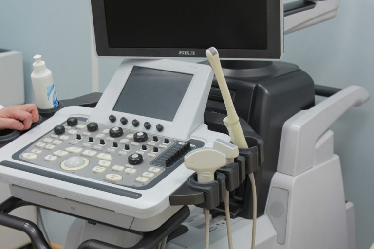
CPAM is most commonly identified during a prenatal ultrasound.
It may be discovered during a routine anatomical scan, often around the middle of pregnancy.
During ultrasound, the doctor may see an abnormal mass within one side of the baby’s chest.
The appearance can vary depending on the type of CPAM.
Macrocystic CPAM
This type contains relatively large fluid-filled cysts that can be visible on ultrasound.
Microcystic CPAM
This type consists of many small cysts and may appear more solid on ultrasound.
Color Doppler can also be useful when evaluating the blood supply to the lesion and distinguishing CPAM from other fetal lung abnormalities.
What Other Conditions Can Look Like CPAM?
Not every fetal chest mass is CPAM.
Other conditions that may have a similar appearance include:
- Bronchopulmonary sequestration
- Hybrid CPAM and sequestration lesions
- Congenital diaphragmatic hernia
- Congenital lobar emphysema
- Other fetal lung lesions
- Rare thoracic masses
Accurate diagnosis is important because different conditions can require different approaches to monitoring and treatment.
Can Fetal MRI Be Used?
Yes.
In selected cases, your healthcare team may recommend fetal magnetic resonance imaging (MRI).
MRI can provide additional information about:
- The location of the lesion
- Its size
- The amount of normal lung remaining
- The effect of the lesion on surrounding structures
- The relationship between the lesion and the diaphragm
MRI may be particularly useful when ultrasound findings are unclear or when the healthcare team needs more information for pregnancy and delivery planning.
Not every fetus with CPAM requires fetal MRI.
What Is the CPAM Volume Ratio?
One important measurement used during prenatal monitoring is the CPAM volume ratio (CVR).
The CVR compares the estimated volume of the lung lesion with the baby’s head circumference.
This measurement helps doctors estimate how large the lesion is relative to the size of the fetus.
A higher CVR can indicate an increased risk of complications, particularly fetal hydrops. A CVR of approximately 1.6 or higher before 26 weeks has been associated with increased risk of hydrops and is one reason that closer fetal monitoring or referral to a fetal treatment center may be considered.
The CVR is only one part of the assessment. Your OB-GYN or maternal-fetal medicine specialist will also consider the baby’s overall condition and ultrasound findings.
Can CPAM Affect the Baby Before Birth?
Many CPAMs remain relatively small and do not cause significant problems during pregnancy.
However, a large lesion can occupy space within the fetal chest and compress the developing normal lung.
In severe cases, the mass can also displace the heart and interfere with circulation.
Potential complications include:
- Compression of normal lung tissue
- Pulmonary hypoplasia
- Heart displacement
- Fetal hydrops
- Abnormal fetal circulation
- Maternal mirror syndrome in severe cases
Large lesions that cause significant pressure within the chest require particularly close monitoring.
What Is Fetal Hydrops?
Fetal hydrops, or hydrops fetalis, occurs when abnormal amounts of fluid accumulate in at least two fetal compartments.
Ultrasound may show fluid:
- Around the heart
- Around the lungs
- In the abdomen
- Under the skin
Hydrops is a serious complication because it can indicate that the fetus is having difficulty maintaining normal circulation.
Large CPAMs can occasionally cause hydrops by compressing the heart and normal lung tissue.
If hydrops develops, management should be coordinated urgently with a specialist fetal care team.
Can CPAM Get Smaller During Pregnancy?
Yes.
This is one of the reasons that serial ultrasound monitoring is important.
A CPAM may appear to grow during the earlier stages of pregnancy, but its growth may slow later in pregnancy while the fetus and normal lung tissue continue to develop.
As a result, the lesion can appear relatively smaller later in pregnancy even if it has not completely disappeared.
Some CPAMs may become difficult to see on later ultrasound examinations. However, this does not necessarily mean that all abnormal tissue has disappeared.
How Is CPAM Managed During Pregnancy?
Management depends on:
- Size of the lesion
- CVR
- Type and appearance of the lesion
- Gestational age
- Fetal lung development
- Presence or absence of hydrops
- Heart function
- Associated abnormalities
Serial Ultrasound Monitoring
Most babies with CPAM are monitored with repeat ultrasound examinations.
Your OB-GYN may monitor:
- Lesion size
- CVR
- Fetal growth
- Heart function
- Amniotic fluid
- Evidence of hydrops
- Development of the normal lungs
The frequency of ultrasound depends on the severity and behavior of the lesion.
Small, stable lesions may require less frequent monitoring, while large or rapidly growing lesions may require very close surveillance.
Fetal Echocardiography
A fetal echocardiogram may be recommended, particularly when the lesion is large or there are concerns about the baby’s heart or circulation.
This allows specialists to evaluate cardiac structure and function.
Does CPAM Require Treatment Before Birth?
Most CPAMs do not require fetal surgery.
If the lesion remains small and the baby shows no evidence of hydrops or significant compression, careful monitoring is usually the primary approach.
Rarely, a large CPAM can become life-threatening before birth. In highly selected cases, fetal intervention may be considered at a specialized fetal treatment center.
Possible fetal interventions include procedures designed to drain large cysts, such as a thoracoamniotic shunt, or other specialized interventions depending on the lesion and clinical circumstances.
These procedures are not routine treatment for CPAM and are generally considered only in severe cases.
Can Steroids Be Used for CPAM?
In selected pregnancies involving a large CPAM and significant risk of hydrops, specialists may consider maternal corticosteroids.
The decision depends on the lesion, gestational age, fetal condition, and specialist assessment.
Steroid treatment should only be given under the supervision of the pregnancy care team.
Does CPAM Mean My Baby Has Other Birth Defects?
Many babies with CPAM have no other major abnormalities.
However, additional congenital abnormalities can occur in a minority of cases. Some sources estimate associated anomalies in approximately 10% to 20% of CPAM cases.
For this reason, a detailed ultrasound examination is important after CPAM is identified.
Your healthcare provider may assess:
- Heart
- Kidneys
- Brain
- Spine
- Abdomen
- Other organs
Is CPAM Associated With Chromosomal Abnormalities?
CPAM itself is not generally considered a chromosomal disorder.
However, because other congenital abnormalities can occasionally occur alongside CPAM, your doctor may discuss whether additional genetic evaluation is appropriate based on the overall ultrasound findings and your pregnancy history.
The decision about genetic testing should be individualized.
Can CPAM Affect Delivery?
In many cases, CPAM does not require an early delivery.
If the baby remains stable and there are no other complications, pregnancy can often continue under appropriate monitoring.
However, delivery planning becomes particularly important when:
- The lesion is large
- There is evidence of fetal hydrops
- The baby has significant lung compression
- There are other fetal abnormalities
- Neonatal respiratory problems are anticipated
When significant respiratory problems are possible, delivery may be planned at a hospital with appropriate NICU, neonatology, pediatric surgery, and respiratory support capabilities.
Is Cesarean Delivery Required for CPAM?
No.
CPAM by itself does not automatically require a cesarean section.
The safest mode of delivery depends on:
- Fetal condition
- Size and severity of the lesion
- Obstetric indications
- Fetal position
- Previous maternal surgery
- Other pregnancy complications
Your OB-GYN and fetal care team will determine the safest delivery plan.
What Happens to a Baby With CPAM After Birth?
The baby’s condition after delivery can vary considerably.
Some babies are completely stable and have no immediate breathing problems.
Others may develop:
- Rapid breathing
- Difficulty breathing
- Low oxygen levels
- Cyanosis
- Respiratory distress
- Pneumothorax
Newborns with significant respiratory symptoms may require immediate neonatal support.
How Is CPAM Confirmed After Birth?
Postnatal imaging is generally performed even when CPAM was identified during pregnancy.
A chest X-ray may be performed initially.
Additional imaging, particularly CT of the chest, may be used to define the lesion and its anatomy.
The pediatric team may also evaluate the baby’s oxygen levels, breathing, and overall condition.
Does CPAM Require Surgery?
Surgery is commonly used to remove the affected portion of the lung, particularly in symptomatic infants.
The operation is often a lobectomy, in which the affected lung lobe is removed.
In selected asymptomatic infants, the decision to operate versus monitor the lesion can be individualized. Evidence and practice vary regarding the management of asymptomatic CPAM, so the pediatric surgeon will consider the size, anatomy, symptoms, infection risk, and other factors.
A 2024 Philippine Children’s Medical Center review reported that lobectomy was the preferred surgical intervention in its series, with generally favorable outcomes.
What Is the Long-Term Outlook for Babies With CPAM?
The prognosis is generally good when the lesion is small and does not cause severe complications before birth.
Babies who require surgery can often go on to live healthy lives.
The lungs also have significant capacity for growth during childhood. Some children who undergo removal of an affected portion of the lung can develop normally as the remaining lung tissue grows.
The outlook depends primarily on:
- Size of the CPAM
- Presence of hydrops
- Amount of normal lung development
- Other congenital abnormalities
- Condition at birth
- Need for neonatal respiratory support
CPAM During Pregnancy in the Philippines
For pregnant women in the Philippines, prenatal detection of CPAM provides an opportunity to establish a coordinated care plan before delivery.
Once CPAM is suspected, an OB-GYN may refer the patient to a maternal-fetal medicine specialist for detailed evaluation.
Depending on the severity, care may involve collaboration among:
- OB-GYN
- Maternal-fetal medicine specialist
- Fetal medicine specialist
- Pediatric radiologist
- Neonatologist
- Pediatric pulmonologist
- Pediatric surgeon
- Anesthesiologist
Because CPAM is uncommon, referral to a tertiary hospital with appropriate neonatal and pediatric surgical facilities may be recommended when the lesion is large or associated with complications.
The Philippine Children’s Medical Center has published local data on CPAM, demonstrating that prenatal ultrasound can identify the condition and that surgical treatment can have favorable outcomes.
Questions to Ask Your OB-GYN After a CPAM Diagnosis
If your baby’s ultrasound shows a possible CPAM, consider asking:
- How large is the CPAM?
- What is the CVR?
- Is the lesion cystic or solid?
- Is the baby’s heart being compressed?
- Is there evidence of hydrops?
- Is the baby growing normally?
- Does my baby need a fetal echocardiogram?
- Would fetal MRI provide additional information?
- How frequently should ultrasound monitoring be performed?
- Where should I deliver?
- Will a neonatologist and pediatric surgeon need to be available at delivery?
- Will my baby require surgery after birth?
Writing down these questions before your appointment can make it easier to understand the treatment plan.
When Should You Seek Urgent Medical Attention?
CPAM itself usually does not cause symptoms in the mother. However, pregnant women should seek prompt medical assessment for symptoms such as:
- Vaginal bleeding
- Leakage of fluid
- Regular contractions before term
- Severe abdominal pain
- Markedly reduced fetal movement
- Severe headache or visual disturbances
- Sudden swelling or other concerning pregnancy symptoms
These symptoms may have causes unrelated to CPAM but should still be evaluated promptly.
Final Thoughts
Congenital Pulmonary Airway Malformation is a rare developmental abnormality of the fetal lung that is increasingly diagnosed during routine prenatal ultrasound. Although hearing that your baby has CPAM can be frightening, many cases are small, remain stable, and have good outcomes.
The most important factors are the size and behavior of the lesion, the development of the baby’s normal lungs, and whether complications such as fetal hydrops develop.
For most pregnancies, the main approach is careful ultrasound monitoring and appropriate delivery planning. Severe cases may require referral to a specialized fetal care center and, rarely, intervention before birth.
If your baby has been diagnosed with CPAM in the Philippines, an experienced OB-GYN or maternal-fetal medicine specialist can help you understand the ultrasound findings, arrange appropriate monitoring, and coordinate care with neonatal and pediatric specialists when necessary.